Document Actions
Imaging Colloidal Suspensions
Traditionally colloids are studied using light scattering (static and dynamic). Recent advances in optical microscopy, however, have made available a host of real-space methods for direct observation of suspensions at single-particle level under a variety of conditions. COSMIC provide
a unique concentration of resources for such studies.
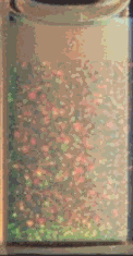
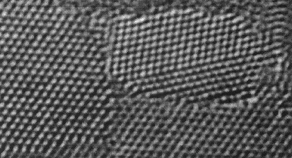
On the left is a sample of colloidal polycrystals as they appear to the naked eye (sample cell 1 cm across). The right hand side shows a differential interference contrast micrograph of a similar sample. The crystalline packing of the (PMMA) colloidal spheres (1 micron diameter) is clearly seen. The iridescent colours in the first sample are due to the Bragg diffraction of incident white light.
Projects in COSMIC will include
- Colloidal gels — particles can, under suitable conditions, be induced to aggregate to form open, space-spanning structures (somewhat like Swiss chess!) know as gels. The evolution of these structures with time can be subtle, but can be followed in detailed using confocal microscopy. Also, the way such structures resist stress is largely unknown. Optical tweezers should allow us to perturb gels locally and measure the forces holding them together. Part of this project is Colloidal Gels Under Flow.
- Nucleation — how does crystalline order emerge from a disordered, fluid-like arrangement of particles? The fast, Nipkov disc confocal microscope should allow us to follow this process with unprecedented detail. Of particular interest will be crystals formed from suspensions with two different sizes of particles.
- Jamming — when a suspension is subjected to fast shear, it may ‘jam’ and turn into something like a solid. One theory suggests that there are ‘force chains’ of particles jammed together in a specific way to resist the applied shear. The close approach of the surfaces of particles in force chains should be susceptible to direct observation (for the first time) using fluorescence lifetime imaging microscopy (FLIM). At the same time, the stresses developed in such chains (if they exist!) can be mapped using optical tweezers.
- Templated growth — we want to develop programmable multiple tweezers in order to grab particles in a particular spatial configuration. This produces an ‘artificial nucleation’ upon which crystal growth may develop, giving structures otherwise not accessible through equilibrium processes alone.
- Direct measurement of fluorescence energy transfer — the fluorescence resonance energy transfer (FRET) between donor and acceptor fluorophores is supposed to depend on the inverse sixth power of the distance between molecules. Using optical tweezers to hold two particles coated with donors and acceptors respectively, we should be able to measure this dependence directly using FLIM. The results should be of significance to biologists, who use FRET heavily as a ‘distance ruler’.